Welcome back to another edition of: “Who Wants to be a Plant
Diagnostician?” with your hosts, the Jefferson County Plant Diagnostic Clinic.
This week, we wanted to focus on the mantra, “Test, Don’t Guess!”, as it
applies to plant diagnostics. In the world of plants, there are many pathologies
that present similarly, so it’s critical to thoroughly evaluate the problem at hand and the possible causes in order to best create a management plan. Below, we’ve put
together a few examples of some lookalikes that have recently come into our
clinic…
Test, Don’t Guess!
***
When you hear the words “spots” and “tomatoes” in the same sentence, you may automatically assume tomato spotted wilt virus (TSWV), a virus vectored by adult thrips. And in some cases, you may be correct. For example, we received a recent inquiry regarding tomatoes presenting with chlorotic rings (shown below), a classic indicator of TSWV:

Tomatoes believed to have tomato spotted wilt virus (TSWV).
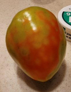
However, this assumption does not always hold up. In another recent tomato inquiry, we received photos of tomatoes that also have yellow spots (shown below); these spots, we think, are caused by stink bug feeding. Stink bugs typically feed on green fruit and produce dark pinpricks, surrounded by white tissue. However, as the fruit ripens, these damaged areas turn yellow.


Tomatoes with spots believed to be
from stinkbug feeding.
We say “we believe” the first
example of tomatoes has TSWV and the second example was fed on by stink bugs
because we haven’t received physical samples to the clinic yet. These are our
initial suspicions and, to confirm, we need to TEST! To differentiate between TSWV
and stink bug feeding, we would first evaluate the plant for other symptoms. TSWV
may result in bronzing leaves, leaves with small, dark spots, tip dieback, streaked
terminal stems, or drooping leaves. Conversely, stink bug damage should be confined
to the fruit itself. At the end of the day, symptoms may be variable for TSWV
though, so, fortunately, we have a great tool at our clinic to rule-out TSWV: a
TSWV antibody test! As TSWV and stink bugs have very different implications for
management and the fate of the plants, TESTING, and not GUESSING, is key!
***
Switching over now to woody plants, we received an inquiry regarding spruce trees that were experiencing browning/purpling of needles (see below) and extensive interior needle drop. Due to the cultural characteristics of the affected trees (very mature, very closely-spaced together, located near greater exposure to temperature extremes and winds, etc.) and the purpling of the needles, we felt confident that the trees were experiencing a cycle of high competition for water, drought stress, and winter dessication. However, due to the browning of the needles, needle shedding, and dark banding observed on some needles (see second image below), there was concern regarding fungal colonization of the spruce (i.e. needle cast fungi).


Discoloration and dark bands found on spruce, likely due to water stress and winter desiccation.
Although needle cast diseases are
rarely found in dry environments, we made sure to TEST, not GUESS! Roughly one
week after plating a number of suspicious areas of needle discoloration onto growth
media, we had several different-looking fungal growths on our plates! In examining
these various fungal growths under the microscope, we found no evidence of
needle cast fungi; rather, we saw several types of saprophytic fungi (see example below) that are typically found in our environment and would not cause damage
to spruce. Better to be safe than sorry!

Hyphae of saprophytic fungi (consistent
with Alternaria/Ulocladium).
***
Our last example of the day deals
with another woody plant: the Austrian pine. We’ve had a handful of individuals
bring samples into the clinic lately, all highly concerned about Pine Wilt/Pinewood
Nematode (PWN). The trees generally present with needle discoloration and
rapid dieback or death of the tree, which is consistent with PWN. Additionally,
some of the samples reveal bluestain fungus in the wood (shown below), which is
another indicator of PWN. However, none of these symptoms are conclusive
of PWN, so we always have to...you guessed it...test, not guess!

Blue stain found on Austrian pine.
Further examination
of the specific sample shown above revealed exit holes, galleries, and Ips pini beetles
(see below). Egg galleries of Ips are usually Y- or H-shaped; upon
hatching, larvae create smaller lateral galleries from the main gallery. Due to
Ips tunneling, affected parts of the tree discolor and die, consistent
with the symptoms of our samples. However, Ips typically attack already
stressed and weakened trees, so while we have clear evidence of Ips, we
can’t rule out PWN without a further test!


Exit holes (top-right of top image)
and galleries/frass (both images) made by Ips pini, found in Austrian pine.
By running a PWN-specific test, we can extract and identify the
nematodes responsible for Pine Wilt Disease, if present. Most of our samples
came back negative for PWN; however, we did find a few nematodes in the sample (shown
below)! BUT, they don’t have the same physical features as PWN…so, on to
another test: we’re working through the identification of these nematodes to
identify whether they are the Pine Wilt Nematode, Bursephalenchus xylophilus,
or one of the many non-pathogenic nematodes that live in and around the tree.
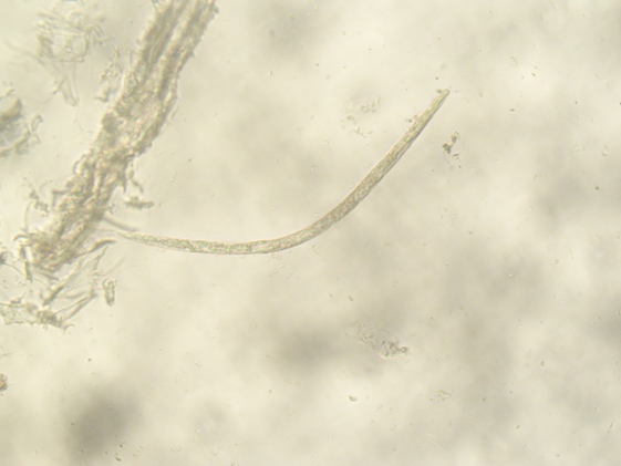
Our unidentified nematode, approximately
300-350 microns in length.

This is an adult, male Pine Wilt
Nematode; length approximately 1000 microns. Photo Credit: Pest and Diseases
Image Library, Bugwood.org.
And that concludes today's edition of "Who Wants to be a Plant Diagnostician"! While I've most certainly overused this phrase throughout the course of this blogpost, I'm going to reiterate it one more time...TEST, DON'T GUESS!!!
No comments:
Post a Comment